
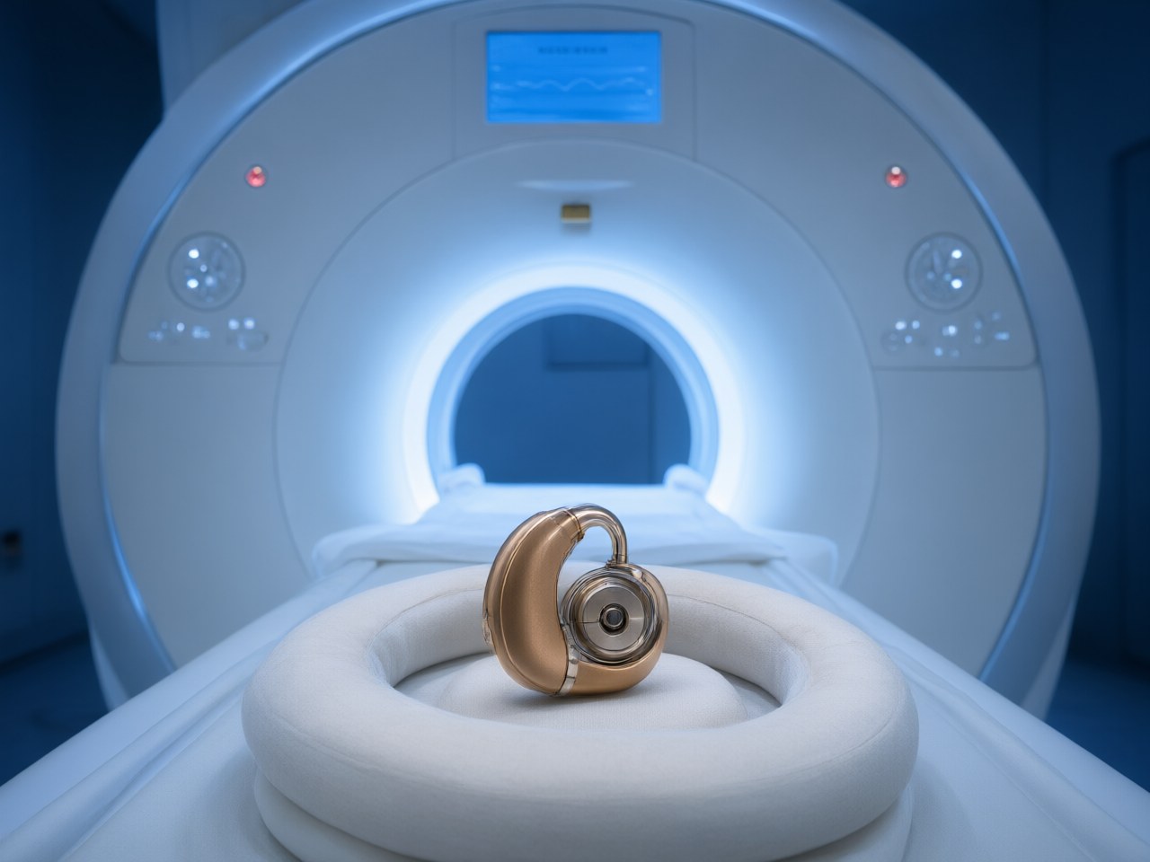
A healthy volunteer study reveals that scanning with a 0.55-tesla mid-field magnetic resonance imaging scanner substantially reduces cochlear implant magnet displacement compared to standard 1.5-tesla systems while preserving clinically acceptable temporal bone imaging. The…
Republished by AIC Engineering. All rights belong to the original publisher; see Source below.
A healthy volunteer study reveals that scanning with a 0.55-tesla mid-field magnetic resonance imaging scanner substantially reduces cochlear implant magnet displacement compared to standard 1.5-tesla systems while preserving clinically acceptable temporal bone imaging. The researchers hypothesised that 0.55T MRI would reduce magnet displacement risk and pressure perception compared to 1.5T MRI in volunteers with bandaged cochlear implants while maintaining temporal bone image interpretability.
To rigorously test this hypothesis, the investigators designed a prospective experimental protocol evaluating ten healthy volunteers who were bandaged with externalized cochlear implants. Five participants were fitted with devices utilizing conventional axial magnets, while the remaining five participants received devices containing rotating diametric magnets. Each volunteer underwent consecutive 0.55T and 1.5T MRI sessions.
Measurements revealed that the axial magnets experienced a torque of 0.133 Newton-meters within the 1.5T scanner compared to 0.062 Newton-meters within the 0.55T system. Consequently, axial magnets dislodged in four out of the five volunteers following their 1.5T imaging session, whereas zero displacement events occurred during mid-field 0.55T scans, establishing a statistically significant reduction in dislocation risk with a p-value of 0.04. Conversely, rotating diametric magnets resisted displacement across both magnetic field strengths, displaying closely matched torque values of 0.046 Newton-meters at 1.5T and 0.044 Newton-meters at 0.55T.
Alongside physical displacement tracking, the researchers assessed subjective pressure perception among all ten participants using a standardized visual analogue scale. Pressure perception did not differ between the 0.55T and 1.5T MRI systems.
The study also comprehensively evaluated temporal bone image interpretability across seven participants scanned using two dedicated protocols: two-dimensional T2-weighted fast spin echo sequences and constructive interference in steady state sequences. Evaluating clinicians analyzed overall diagnostic confidence, the extent of image degradation caused by implant-related metal artifacts, and the physical distances separating artifacts from key anatomical landmarks. Both imaging sequences provided adequate diagnostic confidence across both magnetic field environments, confirming that mid-field acquisition protocols maintained diagnostic interpretability despite operating at a noticeably lower baseline magnetic field strength.
Detailed comparative analysis showed that the 0.55T two-dimensional T2-weighted fast spin echo sequence actively reduced cochlear implant-related image artifacts relative to higher-field acquisitions while enabling a diagnostic confidence score comparable to 1.5T scans. Furthermore, statistical analysis using paired Wilcoxon signed-rank tests for ordinal diagnostic parameters and Bland鈥揂ltman plots for anatomical distances demonstrated that the spatial boundaries separating device-induced artifacts from native temporal bone structures did not significantly differ between scanner types.
In their clinical appraisal, the authors noted that mid-field 0.55T magnetic resonance imaging mitigates axial cochlear implant magnet displacement without compromising necessary anatomical visualization of the temporal bone. The researchers concluded that mid-field imaging could provide a viable, clinically valuable option for individuals fitted with axial magnet implants, particularly those who have previously suffered magnet dislodgement, severe pain, or complications during conventional high-field diagnostic imaging.
Source
- Bioengineer.org (2026-11-12)
- Original article: Mid-Field MRI Minimizes Cochlear Implant Magnet Displacement in Volunteer Study
- https://bioengineer.org/mid-field-mri-minimizes-cochlear-implant-magnet-displacement-in-volunteer-study/